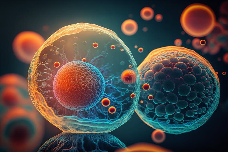
A Microscope’s View of Stem Cells: What We’re Learning
Why Seeing Is Believing
When you pull a slide under a high‑resolution lens, the abstract notion of a stem cell suddenly gains texture. You can watch a tiny nucleus pulse with activity, spot the faint glow of a fluorescent tag, or trace the delicate web of a colony expanding across the dish. Those visual clues translate into the big questions researchers ask every day: how do these cells decide whether to become a neuron, a muscle fiber, or stay in a pluripotent state?
The Main Tools in the Lab
Modern microscopes are far from the simple light boxes of a century ago. A typical stem‑cell lab might rotate among three workhorses:
- Bright‑field microscopy – the classic, low‑cost option that lets you gauge colony size and overall morphology.
- Fluorescence microscopy – by tagging proteins with fluorescent dyes, scientists can pinpoint where key regulators sit inside the cell.
- Confocal or multiphoton systems – these slice through thick specimens, offering three‑dimensional reconstructions of cellular neighborhoods.
Each method paints a slightly different picture, and the best insights usually come from layering them together.
Bright‑field basics
With bright‑field, the cell looks like a translucent grain of sand on a watery background. Researchers count the number of colonies, note edge smoothness, and watch for any rogue differentiating cells that break the uniform look. Though the image is simple, it tells you whether your culture conditions are stable enough to keep the cells in their stem state.
Fluorescent fingerprints
Fluorescent markers are the real game‑changers. For example, an antibody linked to a green dye might bind OCT4, a protein that signals pluripotency. When the microscope lights up, the nucleus glows green if OCT4 is present. A red‑tagged antibody could highlight SOX2, another pluripotency factor. By overlaying these colors, you can tell at a glance whether a cell is still “stem‑like” or has started to specialize.
Peeking into depth with confocal
Confocal microscopes bounce a laser through the sample and capture light only from a thin focal plane. The result is a crisp slice, free of out‑of‑focus blur. Stack enough slices together and you get a 3‑D model of a stem‑cell niche—a miniature ecosystem where neighboring cells send signals that guide fate decisions. This depth perception is essential when studying organoids, those mini‑organs grown from stem cells.
What the Images Reveal About Stem‑Cell Behavior
Beyond pretty pictures, microscopy provides concrete data. Researchers measure:
- The intensity of fluorescent signals, which correlates with protein expression levels.
- The spatial relationship between nuclei and cytoplasmic markers, hinting at signaling pathways in action.
- The dynamics of cell division, captured through time‑lapse imaging that shows how often a stem cell splits and whether its daughters remain identical.
These observations feed into models that predict how a batch of stem cells will behave when transplanted into a patient or when coaxed into forming a specific tissue type.
Common Pitfalls and How to Avoid Them
Even the best microscopes can mislead if the preparation isn’t right. Here are a few practical tips:
- Fixation timing: Over‑fixing can mask antigens, making fluorescent tags appear dim.
- Background fluorescence: Unbound dye can create a haze; thorough washing reduces this noise.
- Photobleaching: Prolonged exposure to the laser can fade the signal. Using anti‑fade reagents or limiting exposure time helps preserve brightness.
Attention to these details keeps the visual data reliable and reproducible.
Future Directions: Live‑Cell Imaging and Beyond
One exciting frontier is watching stem cells in real time without killing them. Advances in low‑toxicity dyes and adaptive optics now let scientists record the same cell’s journey from pluripotency to a specialized fate over days. Pairing these videos with AI‑driven analysis may soon reveal subtle patterns—like micro‑oscillations in gene expression—that were invisible to the naked eye.
Quick FAQ
What does “pluripotent” mean under the microscope?
Pluripotent cells typically show a large, round nucleus with a high nucleus‑to‑cytoplasm ratio and strong expression of markers like OCT4 and SOX2, which appear as bright fluorescent spots.
Can I use a regular light microscope to study stem cells?
You can get basic information about colony size and shape, but identifying specific proteins or tracking intracellular processes generally requires fluorescence or confocal imaging.
How often should I image my stem‑cell cultures?
For routine quality checks, a snapshot every few days suffices. For experiments tracking differentiation, time‑lapse imaging every 10‑20 minutes over several hours provides useful kinetic data.